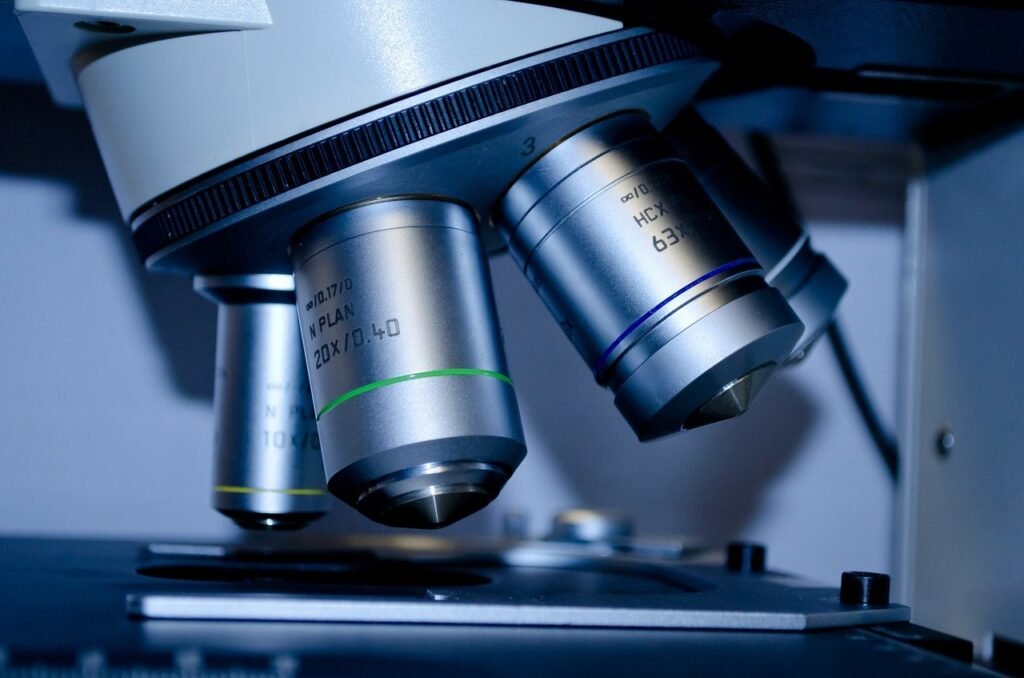
There was no easy way to share what you saw under a microscope because it required someone else to peek into the instrument physically. People gradually realized that the effects of a diagnosis, whether histopathology, hematology, cytology, microbiology, or other applications, needed to be recorded.
The only way to maintain records and share images is to capture them with a suitable camera. This is how the idea of a digital camera for microscope eyepiece came into being. High-quality images captured from pathology samples helped transform medical practices. Patients and doctors could now use them for second opinions and tumor boards. Printing out images eliminated the need to carry or ship slides for diagnosis. The digital images also add value to the information used for publications and teaching. But choosing the best microscope eyepiece camera becomes daunting with the plethora of options available. Each one comes with an array of specifications and technical details, making you question your choice. To pick the most appropriate camera that will cater to your needs, simply ask yourself a few questions.
1) What Contrast Techniques You Will Be Needing?
There are a few guiding principles to start with.
If your profession requires you to capture only brightfield images, a high-quality color camera would be appropriate.
If you want to capture fluorescent images occasionally, SCMOS cameras come in handy, but they are not suitable for long-exposure applications, like capturing bioluminescent images.
There is also a decision to be made whether you require a monochrome or colored camera. Monochrome cameras are preferred while working with fluorescent-labeled samples.
2) What Is the Level of Resolution Needed?
In microscopy, the features required for a digital camera for a microscope eyepiece are quite distinct from what you would look for in a consumer camera. Choosing a camera with higher megapixels will not be enough to satisfy your needs. Some factors like chip size, frame rate, and pixel size need to be considered.
Lower magnification – if your purpose is to capture images of cells and tissues in routine applications, magnifying them up to 40x in brightfield, the recommendation would be at least a three-megapixel camera chip. Any medium-range pixel size should be enough to deliver the image quality you want. A cooling feature is not mandatory.
Highest magnification – scientists looking to research the details of nuclei in 100x oil, studying hematology, or observing the movements of bacteria in microbiology, need high-end resolution. The best combination that can capture the truest image is a camera with optimized pixel size and a decent number of megapixels.
Live image – live images are increasing in importance for teaching and tumor board discussions. They can be projected on a monitor with or without a computer. But for a smooth visual of the microscopic image, the camera needs to have a high frame rate (fps or frames per second). Technological advancement has further made it easier to directly connect the cameras to a screen without a computer.
3) Which Interface Will You Be Comfortable With?
A camera eyepiece microscope can be connected to computers via USB 2.0, USB 3.0, or firewire. It is important to figure out whether your computer has matching sockets or can be upgraded. Only a high-efficiency computer will be compatible to cope with imaging tasks.
If you need live camera images frequently for teaching or discussions that need to be projected directly on a screen, your ideal choice would be a camera with an HDMI connection. They are easy to connect to monitors. Also, look out for cameras with high fps to play the live images smoothly.
Some people do not want to be bothered to connect to a computer every time they are using a digital eyepiece for the microscope. They opt for cameras with an sd card slot to keep the images saved.
4) What Technical Details to Look for in a Camera Chip?
Is fast frame rate (CMOS) what you are looking for or high sensitivity (CCD)? CMOS (complementary metal-oxide-semiconductor) and CCD (charge-coupled device) are the two terms commonly heard while looking out for chips in microscopy cameras. They have their own advantages and disadvantages. In the past, CMOS sensors were not a frequent choice as they failed to efficiently convert the incoming light into electrical signals. So, people moved on to CCD sensors, which worked excellently for low-light applications. However, it was soon realized that CMOS chips allowed a higher frame rate, making them perfect for live images.
The wide variety of choices with complex, interrelated key elements often tends to baffle consumers. It is best to base the decision on the requirements for your microscopic observation. The ever-advancing technology serves higher image quality and appropriate image processing that has enabled researchers to break through the traditional limitations of microscopic imaging and discover the unknown.
People Usually Search Keywords: Microscope Manufacturers | Laboratory Microscope | Microscope Manufacturers in India | Microscope Supplier | Microscope Suppliers in India | Laboratory Microscope Suppliers | Microscope Manufacturers in Ambala | Microscope India | Best Microscope Manufacturers | Microscope Ambala | Microscope Online India | Microscope Brands in India | Microscope Companies in India | Microscope Online Shopping India | Top Microscope Brands in India | Indian Microscopes | Microscope India Suppliers | Top Microscope Manufacturers | Best Microscope Brands | Best Microscope Companies | Microscope Brands | Microscope Companies | Microscope Vendors
What Is a Binocular | Binocular Microscopy | Binocular Microscope | Binocular Microscopes | Binoculars Microscope | Microscope Binocular | Uses of Binocular Microscope | Binocular Microscope Principle | Microscope Comments | Microscope Definition Biology | Define Binocular | What Is Binocular
Uses of Microscope | What Is Microscope | 10 Uses of Microscope | Uses of Lenses in Our Daily Life | Explain Microscope | Define Microscope | 5 Uses of a Microscope | Convex Lens Uses in Daily Life | What Are Its Uses | Purpose of Microscope | Application of Simple Microscope | Use of Simple Microscope | Uses of Simple Microscope | Use of Microscope | Application of Microscope | Uses of Lens in Daily Life
Who Discovered Microscope | Who Invented Microscope | Who Invented the Microscope in 1666 | Zacharias Janssen Microscope | Who Discover Microscope | Who Made the Microscope | Microscope Invented by | Who Invented Compound Microscope | Who Invented the Microscope and When | Microscope Inventor Name | Who Founded Microscope | Microscope in Kannada
What Is a Compound Microscope | Compound Microscope Comments | Compound Microscope Information | Compound Microscope | Compound Microscope Explanation | Discovery of Compound Microscope | Why Is Light Microscope Called a Compound Microscope | Types of Compound Microscope | Compound Microscope Practical | Describe Compound Microscope | About Compound Microscope | Which Lens Is Used in Compound Microscope | Comments on Compound Microscope | Components of Compound Microscope | Explain Compound Microscope | History of Compound Microscope | Study of Compound Microscope


