The Micron Optik Surgical Microscope Neurosurgery configuration is calibrated for the extended working distances and prolonged procedure times typical of complex cranial surgery.
Objective Lens Options: 350mm, 400mm, 450mm, and 500mm focal lengths with enhanced light transmission coatings for deep cavity work.
Focusing Mechanism: Motorized fine focus with 70mm travel range and 1 micron precision plus 350mm coarse focus via column movement.
Fluorescence Channels: Dual fluorescence capability (ICG angiography and 5-ALA tumor detection) with separate foot pedal controls for each modality.
Illumination Delivery: 300W xenon with dual fiber-optic cables providing 200,000 Lux measured at 450mm working distance.
Arm Reach: 1700mm horizontal extension from stand column with 360-degree rotation at every articulation point.
Every neurosurgery-configured surgical microscope includes fluorescence calibration targets and working distance validation certificates.
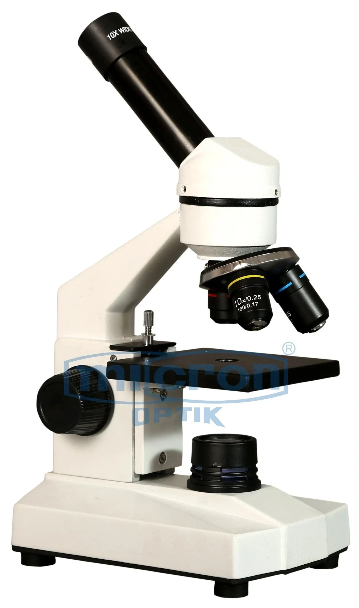
The Surgical Microscope Neurosurgery configuration from Micron Optik represents a purpose-built visualization solution developed in direct consultation with practicing neurosurgeons from India's leading academic medical centers. As a Top Rated and Best Manufacturer with an ISO-certified facility established in Ambala, India since 1966, we have translated specific cranial and spinal surgery requirements into engineering specifications that address real operating room challenges rather than theoretical optical benchmarks. This system prioritizes deep cavity illumination, extended reach, and absolute stability—the three attributes that neurosurgeons consistently identify as most critical for their work. **The microscope has been validated in clinical use at institutions including HBT Medical College, Jjm Medical College Karnataka, and Dr. Ulhas Patil Medical College & Hospital.
The technical foundation of this Surgical Microscope Neurosurgery configuration is the enhanced illumination pathway, which delivers 200,000 Lux of shadow-reduced light through a 450mm working distance without generating significant heat at the surgical field. Traditional microscopes lose 40% to 60% of light intensity over extended working distances, but our dual-fiber delivery system maintains consistent brightness from dura to deep tumor bed. We have harnessed our in-house optical fabrication capabilities to produce objective lenses with enhanced light-gathering characteristics specifically for the 400mm to 500mm working distances commonly used in transsphenoidal and far-lateral approaches. The result is a surgical field that remains brilliantly illuminated even when operating through a narrow tubular retractor at the skull base.
The mechanical architecture of this Surgical Microscope Neurosurgery configuration addresses the spatial constraints of the neurosurgical operating room. The five-point articulated arm with 1700mm horizontal reach allows the microscope to be positioned at the head of the bed while the stand remains outside the anesthesia zone near the patient's waist. Our brand has earned customer loyalty across India because neurosurgeons appreciate that this microscope does not require repositioning between the craniotomy and dural opening phases—the arm geometry maintains reach even as the surgical angle changes from vertical to steep oblique. The 350mm vertical focusing range accommodates patient positioning from supine to lateral to prone without column adjustments.
Choosing Micron Optik as your Manufacturer and Supplier for a neurosurgery-configured surgical microscope means acquiring a system designed for the specific ergonomic demands of cranial and spinal microsurgery. The surgeon-controlled interface places all critical functions on the wireless foot pedal, allowing magnification changes, focus adjustments, and filter selection without looking away from the operative field or removing instruments from the surgical cavity. The documentation system captures both white-light and fluorescence video simultaneously, creating teaching files that show tumor fluorescence before resection and perfusion angiography after vessel reconstruction. Whether you perform 50 or 500 cranial procedures annually, this surgical microscope neurosurgery configuration adapts to your technique rather than forcing you to adapt to the microscope.
This Surgical Microscope Neurosurgery configuration includes features selected specifically for the workflow demands of cranial and spinal procedures.
Fluorescence Intensity Histogram Display: Real-time graphical overlay showing ICG intensity curves allowing quantitative assessment of flow before and after bypass anastomosis.
Motorized Fine Focus with Depth Memory: Programmable focus positions for dura, tumor surface, and deep margin enabling rapid return to previously identified planes after instrument changes.
Sterile Drape Integration Port: Direct drape attachment interface with optical-grade window maintaining sterility without image degradation or reflection artifacts.
Anti-Fog Objective Lens Heating: Resistive heating element embedded in lens housing preventing fog formation when moving from cold to warm operating rooms or during prolonged procedures.
Surgeon Control Preference Storage: User profiles for up to 5 surgeons storing light intensity, magnification limits, focus speed, and filter preferences for instant recall.
These neurosurgery-specific features reduce cognitive load during complex procedures, allowing the surgeon to focus on anatomy rather than equipment management. Contact Micron Optik, the Top Rated and Best Manufacturer and Supplier for specialized surgical microscope neurosurgery configurations in India, to request a technical consultation with our neurosurgery applications specialist.
This Surgical Microscope Neurosurgery configuration supports cranial, spinal, and peripheral nerve procedures with specific optical requirements.
Transsphenoidal Pituitary Surgery: Steep angle capability (0 to 30 degrees tilt) providing visualization through the nostril corridor while maintaining working clearance for endoscopic assistance.
Retrosigmoid Approach (Acoustic Neuroma): Extended working distance (450mm) allowing the microscope to remain outside the surgical field while visualizing the cerebellopontine angle.
Arteriovenous Malformation (AVM) Resection: ICG fluorescence angiography without switching optical paths enabling flow assessment before and after feeder ligation.
Intramedullary Spinal Cord Tumor Removal: Green filter enhancement of interface between tumor and spinal cord parenchyma reducing risk of post-operative neurological deficit.
Peripheral Nerve Sheath Tumor Resection: High magnification (20x to 25x) for fascicular dissection preserving motor fibers while removing schwannomas from major nerve trunks.
Across these neurosurgical applications, this microscope configuration provides the specialized visualization that general-purpose surgical microscopes cannot match.