PHASE CONTRAST ATTACHMENT
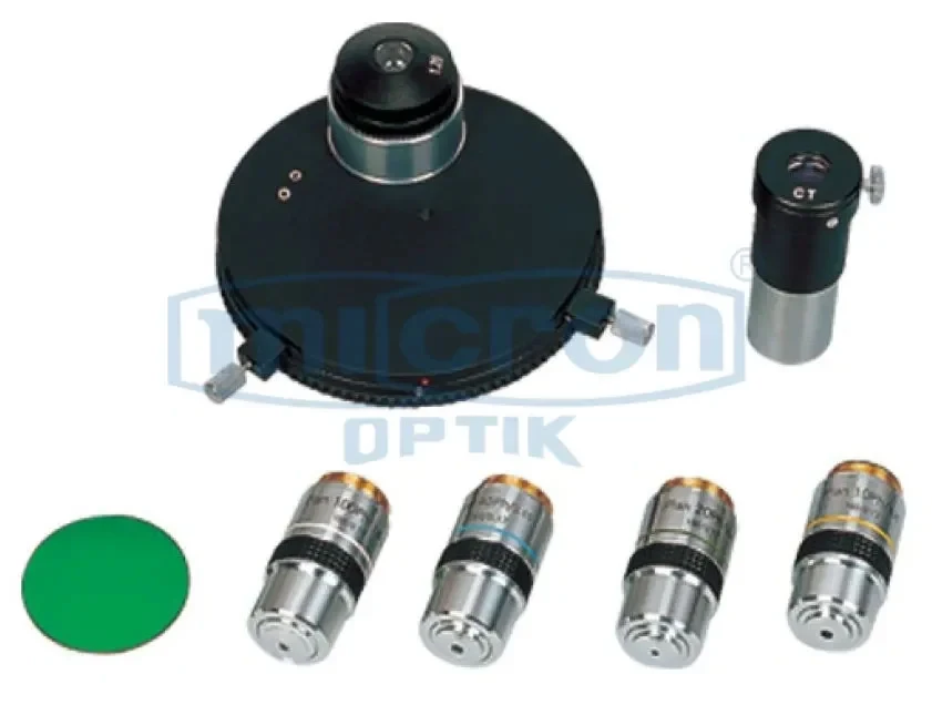
Detailed Phase Contrast Attachment Specifications: Type: Positive phase contrast (most common) or negative phase contrast. Components: Phase condenser (turret or slider type) with annulus plates for different magnifications (e.g., 10X, 20X, 40X, 100X). Phase Objectives: A set of objectives (e.g., 10X, 20X, 40X, 100X oil) with a built-in phase ring. Centering Telescope: A special eyepiece used to align the condenser annulus with the objective's phase ring. Compatibility: Designed for specific microscope models (e.g., Cresta, INV series). Condenser Mount: Fits into the standard substage condenser mount. Aperture: Phase annuli are matched to the N.A. of the corresponding objectives. Illumination: Requires a bright, even light source (halogen or LED). Included: Phase condenser, set of phase objectives, centering telescope, and instructions.
Micron's Phase Contrast Attachment is an advanced optical system that converts a standard brightfield microscope into a powerful tool for observing transparent, unstained specimens. Phase contrast works by translating minute differences in the refractive index and thickness of a specimen into visible differences in brightness. This technique is widely regarded as one of the best methods for observing living cells and other transparent biological materials without staining.
The attachment typically consists of two main components: a specialized phase condenser containing annulus plates, and a set of phase objectives containing a phase ring. When aligned, these components interfere with the light passing through the specimen, creating a high-contrast image. This is one of the best investments for a life science laboratory, as it dramatically expands the microscope's utility. Phase contrast is essential for observing live cell cultures, examining unstained tissue sections, and studying dynamic processes in living specimens. Micron's phase contrast attachment is designed to be easily installed on compatible Micron microscopes, providing a significant upgrade in imaging capability. For clinical labs, research facilities, and teaching hospitals, this is a top rated accessory for live-cell observation.
Designed for precision and consistent performance, Micron's Phase Contrast Attachment enables researchers, laboratory professionals, and educators to examine transparent biological samples with remarkable clarity and contrast. It is especially useful in applications where staining is not possible or where preserving the natural state of the specimen is essential. By enhancing subtle optical differences within the sample, this attachment allows users to observe cell membranes, nuclei, vacuoles, and internal cellular activity that may remain nearly invisible under standard brightfield illumination.
Its robust optical construction and carefully aligned phase elements ensure stable imaging results across routine laboratory use, educational demonstrations, and advanced research workflows. The attachment is ideal for cell biology, microbiology, pathology, and biomedical teaching environments where high-detail observation of live material is required. This makes it one of the best practical upgrades for laboratories seeking improved imaging performance without replacing their entire microscope system.
Micron's Phase Contrast Attachment also supports the study of cell division, motility, cytoplasmic streaming, and other dynamic biological processes in real time. This makes it highly valuable for both teaching and professional diagnostics, where understanding live specimen behavior is critical. It offers a highly effective solution for visualizing delicate structures while minimizing specimen preparation time and preserving sample integrity.
Live Cell Observation Without Staining: Allows for the detailed observation of living, unstained cells and transparent biological specimens by converting phase shifts into visible contrast.
Complete Condenser and Objective Set: The attachment includes a phase condenser with annuli and a matched set of phase objectives for 10X, 20X, 40X, and 100X magnifications.
Centering Telescope for Precise Alignment: Includes a special centering telescope eyepiece that allows the user to accurately align the condenser annulus with the objective's phase ring for optimal image quality.
Significant Microscope Upgrade: A cost-effective way to add advanced phase contrast capability to a compatible Micron brightfield microscope, dramatically increasing its versatility.
Essential for Tissue Culture and Cell Biology: An indispensable tool for monitoring live cell cultures, examining cell morphology, and studying cell division without disrupting the culture.
High-Contrast Images of Transparent Specimens: Reveals fine details and internal structures in unstained samples such as living cells, thin tissue sections, and microorganisms that are invisible in standard brightfield.
Easy Installation on Compatible Microscopes: Designed for straightforward installation on specific Micron microscope frames (e.g., Cresta, INV series), making it a practical upgrade.
Durable and Precision-Made Components: The condenser and objectives are manufactured to high optical and mechanical standards, ensuring reliable, long-lasting performance.
Live Cell Culture Monitoring: Essential for tissue culture labs to non-invasively monitor cell health, confluency, and morphology without staining or removing cells from the incubator.
Cellular and Molecular Biology Research: Used to study dynamic cellular processes such as cell division (mitosis/meiosis), phagocytosis, organelle movement, and cell migration in real-time.
Clinical Diagnostics and Hematology: Applied for examining unstained blood smears, urine sediment, and other body fluids to identify cells, crystals, and casts.
In Vitro Fertilization (IVF) Labs: Critical for assessing the quality and morphology of eggs and embryos without using toxic stains that could harm them.
Microbiology and Parasitology: Used to observe live, unstained microorganisms such as protozoa, algae, and fungi, revealing their natural movement and behavior.
Neuroscience Research: Ideal for examining live neurons and brain tissue slices, allowing researchers to study neuronal structure and network activity.
Pharmaceutical Drug Discovery: Employed in high-content screening assays to assess the effects of drug candidates on live cell morphology and health in a label-free manner.