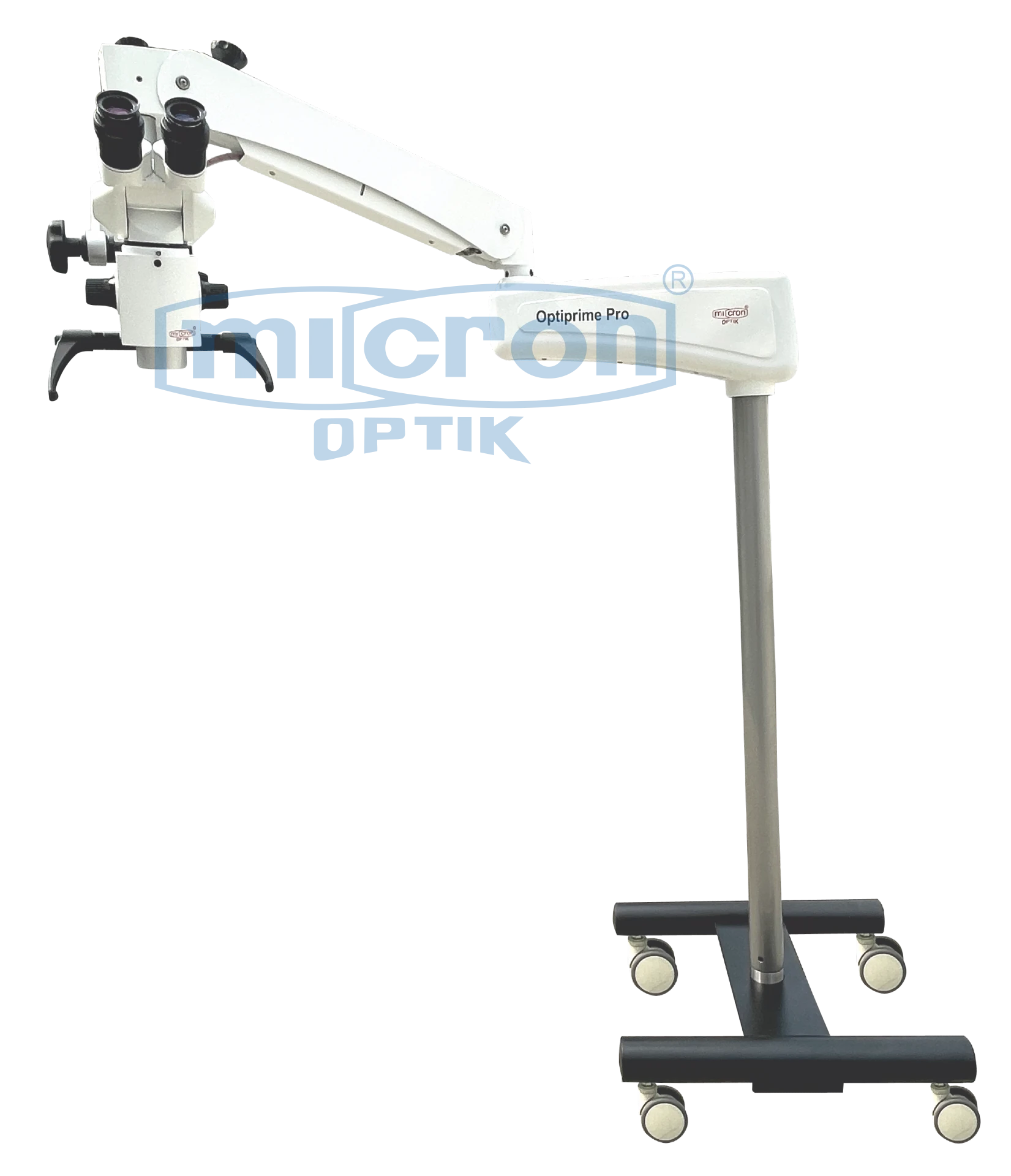
High-resolution apochromatic optical system for true-to-life neural visualization
5-step magnification changer for rapid and precise magnification selection
Wide magnification range of approximately 4× to 25×
High-intensity coaxial LED illumination system
Shadow-free, deep-cavity illumination for cranial and spinal fields
Built-in Green and Orange filters for enhanced tissue and vessel contrast
Interchangeable F = 250 mm and F = 400 mm objective lenses
Ergonomic binocular observation tube optimized for neurosurgical posture
Wide-field, high-eyepoint eyepieces for prolonged microsurgical procedures
Ultra-fine, smooth focusing mechanism for sub-millimeter precision
Heavy-duty mobile floor stand with high stability and lockable casters
Ergonomic handgrips for micro-positioning control
Provision for assistant binocular observation tube
Integrated beam splitter for camera and documentation systems
Optional HD/4K digital imaging system with medical-grade monitor
Designed for long-duration neurosurgical procedures in seated or semi-seated position
Specification | Details |
|---|---|
Application | Neurosurgery, Spine Surgery, Neurovascular Surgery, Micro-surgery |
Optical System | High-resolution Apochromatic Optics |
Magnification Changer | 5-Step |
Magnification Range | Approx. 4× – 25× |
Eyepieces | Wide-field, High-eyepoint, 10× |
Binocular Tube | Ergonomic Neurosurgical Binocular Observation Tube |
Interpupillary Distance | Adjustable |
Dioptre Adjustment | Individual adjustment on eyepieces |
Objective Lenses | F = 250 mm & F = 400 mm |
Illumination | High-Intensity Coaxial LED |
Illumination Control | Variable intensity |
Filters | Green & Orange Filters |
Focusing | Ultra-fine Precision Focusing System |
Stand | Heavy-duty Mobile Floor Stand |
Base | High-stability counterbalanced floor base |
Casters | Lockable Medical-grade Heavy-duty Casters |
Positioning | Smooth multi-axis horizontal & vertical movement |
Camera Provision | Beam Splitter / C-Mount Provision |
Assistant Observation | Provision for Assistant Binocular Tube |
Camera | Optional HD / 4K Digital Camera |
Monitor | Optional Medical-grade HD / 4K Monitor |
Power Supply | 220–240 V AC, 50 Hz |
The Micron Optik Neurosurgical Operating Microscope is specially engineered for high-precision neurosurgical applications including cranial surgery, spinal microsurgery, neurovascular procedures, tumor resection, and complex micro-dissection. It integrates superior apochromatic optics, high-intensity coaxial LED illumination, ergonomic surgeon-centric design, and ultra-stable positioning to deliver exceptional clarity, depth perception, and control in delicate neural structures.
The neurosurgical operating microscope delivers:
Ultra-sharp visualization of neural, vascular, and micro-anatomical structures
Exceptional depth of field for deep cranial and spinal cavities
True color reproduction for accurate differentiation of tissue planes
Minimal optical distortion across all magnification levels
High contrast imaging for fine neurovascular dissection
Stable, fatigue-free viewing for long-duration microsurgical procedures
The integrated coaxial LED illumination system provides intense, focused, and shadow-free lighting aligned with the optical axis, ensuring optimal visibility in deep surgical corridors such as cranial cavities and spinal canals.
The system includes Green and Orange filters to enhance contrast between blood vessels, neural tissue, and surrounding structures, improving precision during delicate dissection and hemostasis.
The system is designed specifically for neurosurgical ergonomics, supporting prolonged procedures with minimal surgeon fatigue. The flexible multi-joint arm system allows smooth, vibration-free positioning over the operating field.
High-stability lockable casters ensure absolute steadiness during surgery, while ergonomic handgrips enable precise micro-adjustments of the optical head.
The system supports advanced neurosurgical imaging and documentation requirements:
Real-time surgical field visualization
High-resolution still image capture
HD/4K video recording of procedures
Surgical documentation and case archiving
Teaching and neurosurgical training applications
Telemedicine and remote consultation support
A beam splitter enables seamless camera integration without compromising the primary surgeon’s optical pathway.
Neurosurgical Operating Microscope Head
5-Step Magnification Changer
Main Binocular Tube (Neurosurgical Ergonomic Type)
Wide-field Eyepieces
F=250 mm Objective
F=400 mm Objective
Coaxial LED Illumination System
Green Filter
Orange Filter
Heavy-duty Mobile Floor Stand
Lockable High-stability Casters
Ergonomic Handgrips
Protective Cover
User Manual
Assistant Binocular Tube
Beam Splitter
HD Digital Camera
4K Digital Camera
Medical-grade Surgical Monitor
Video Recording System
Surgical Image Management Software
Foot Control System
Additional Objective Lenses
Cranial Neurosurgery
Spine Surgery
Neurovascular Surgery (Aneurysm, AVM, etc.)
Tumor Resection (Brain & Spinal Cord)
Micro-neurosurgery
Skull Base Surgery
Peripheral Nerve Surgery
Neurosurgical Training & Education