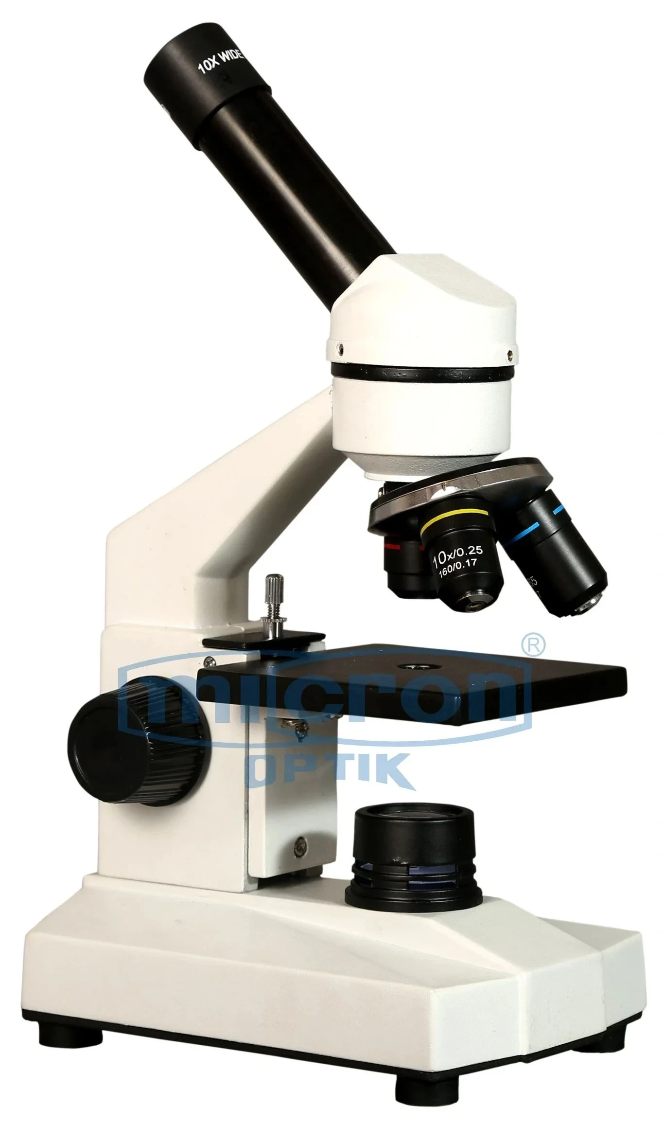
The Micron Optik ENT Microscope integrates precision optics with intelligent mechanical controls for superior otolaryngology outcomes.
Working Distance: 250mm, 300mm, 400mm interchangeable via click-stop objective lenses for ear, nose, and larynx procedures.
Magnification System: Continuous zoom from 3.5x to 25x with parfocal design and 18mm to 5mm field diameter range.
Illumination: 100W LED coaxial illumination with 90,000 Lux intensity, 5700K color temperature, and integrated orange safety filter.
Binocular Head: 0-200° tiltable, 45° inclined tube with 55-76mm interpupillary adjustment and ±6 diopter compensation.
Suspension System: Five-point spring-balanced articulated arm with electromagnetic brakes and 1400mm horizontal reach from stand column.
Every specification is validated through rigorous quality control at our advanced automated facility before dispatch to your hospital.
The ENT Microscope from Micron Optik is a specialized optical instrument engineered to address the unique challenges of ear, nose, and throat microsurgery. As a Top Rated and Best Manufacturer with an ISO-certified facility established in Ambala, India since 1966, we have designed this system to deliver exceptional visualization in the narrow, deep cavities that characterize ENT procedures. It is exceptionally sturdy yet remarkably agile, allowing effortless positioning around the patient's head, shoulders, and upper torso without compromising sterility or workflow. Trusted by leading medical institutions including Grant Govt Medical College, HBT Medical College, and Lokmanya Tilak Municipal Medical College, this microscope sets the benchmark for otolaryngology microsurgery across India.
The defining strength of this ENT Microscope lies in its optimized working distance and coaxial illumination path, specifically calibrated for procedures ranging from myringoplasty to laryngeal microsurgery. The 250mm to 400mm variable working distance provides ample space for micro-instruments, suction tubes, and the surgeon's hands while maintaining parfocal clarity from low magnification for orientation to high magnification for delicate dissection. We have harnessed decades of optical manufacturing expertise to create a system that delivers brilliant, shadow-free illumination exactly where it is needed—deep inside the external auditory canal, the middle ear cleft, or the oropharynx.
Surgeon ergonomics are critically important in ENT, where prolonged procedures with sustained awkward postures can lead to chronic musculoskeletal disorders. The fully articulating suspension arm with electromagnetic brakes allows one-touch positioning, while the 0-200° adjustable binocular tube with 45° inclination enables a natural, upright posture even during complex stapedotomy or cholesteatoma removal. Our brand has earned customer loyalty across India because we understand that a comfortable surgeon is a precise surgeon, and precision in ENT directly correlates with hearing preservation, facial nerve safety, and voice outcomes.
Choosing Micron Optik as your Manufacturer and Supplier means investing in reliability backed by responsive nationwide service and a legacy of excellence since 1966. The ENT Microscope is fully compatible with our high-definition documentation systems, allowing live streaming to teaching theaters, high-resolution recording for medicolegal purposes, and seamless integration with endoscopic and navigation systems. Whether for a new ENT department in a teaching hospital or upgrading an existing otology practice, this microscope delivers the performance, durability, and value that have made us India's most trusted microscope brand. From West Bengal Medical Services Corporation to Department of Health and Family Welfare, our clients consistently choose Micron Optik for their ENT microsurgical needs.
Every feature of this ENT Microscope is purpose-designed for the unique workflow and anatomical challenges of otolaryngology procedures.
Motorized Column with Three Position Memory: Programmable working distance presets for ear (250mm), nose (320mm), and larynx (400mm) allow instant recall with a single foot pedal press.
Integrated Orange & Green Filter Turret: Dedicated orange filter enhances fluorescence detection for cholesteatoma and tumor margins while green filter increases vascular contrast during stapes surgery and tympanic membrane manipulation.
Six-Button Wireless Foot Pedal: Controls magnification, focus, filter selection, video recording, and still image capture without removing hands from micro-instruments or breaking sterility.
Tiltable Objective Lens (0-30° Continuously Variable): Allows visualization around anatomical obstructions like the overhanging tympanic scutum, facial nerve ridge, or middle turbinate.
Integrated 4K Documentation with Direct Live Streaming: Built-in 4K camera with beam splitter captures high-resolution video and images while enabling direct streaming to teaching monitors or remote consultation platforms.
These advanced features collectively reduce operative time, enhance surgical precision, and elevate the educational value of ENT training programs.
From the tympanic membrane to the true vocal cords and beyond, this ENT Microscope enables surgical precision in anatomically challenging regions.
Otologic Surgery (Myringoplasty & Tympanoplasty): Superior coaxial illumination penetrates the external auditory canal revealing the annulus, ossicular chain, and middle ear mucosa with exceptional clarity.
Stapedotomy for Otosclerosis: High magnification (20-25x) and long working distance allow precise fenestration of the stapes footplate and accurate piston placement.
Laryngeal Microsurgery (Vocal Cord Nodules, Polyps & Cysts): Adjustable working distance accommodates suspension laryngoscopes while maintaining focus on vibrating vocal folds for precise phonosurgery.
Cholesteatoma Removal & Ossiculoplasty: Superb depth perception differentiates granulation tissue, keratin debris, and cholesteatoma matrix from healthy mucosa and ossicular remnants.
Rhinologic & Skull Base Procedures: 30° and 45° tilting objective attachments provide visualization around nasal turbinates and into the sphenoid, frontal, and ethmoid sinuses.
Across every ENT subspecialty, this microscope delivers the visualization essential for optimal functional outcomes and patient safety.